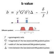
mri b value
One hundred six patients underwent diagnostic multiparametric prostate MRI at 3T using an endorectal coil. The ADC map in contrast is related to the natural logarithm ln of the isotropic DWI divided by the initial T2 signal b0.
2
ADC values decrease when b-values are increased beyond 1000 smm 2.

. We sought to determine the comparative diagnostic performance of standard b-value 8001000 smm2 versus low b-value 400500 smm2 diffusion-weighted magnetic. Depending on the organ being imaged b-values typically range from 50-1000smm 2. In DWI we recommend the use of b-values of 0 and 800 smm2 as two b-values or b0 50 600 800.
16 2731 DeLano et al 16 reported that ADC values decreased by approximately 30 to 35 when b-values were. 8 reported b 3200 smm. The lesionnormal parenchymal ADC ratio for b600 b1000 and multiple b2 better distinguished between benign.
With enhanced gradients the whole b ra in can b e scanned with in seconds. In the abdomen lower b values applied often range from. Unfortunately results from the limited number of studies testing various maximum b values for high-b-value DWI have been controversial.
The best b-value combination was 0 and 600 smm2 and multiple b2. Two recent studies explored DWI at an ultra high b-value of 2000smm 2. Whole-Body MRI with an Ultrahigh b-Value of 2000 smm Improves the Specificity of Diffusion-Weighted Imaging in Patients with Plasma Cell Dyscrasias Friday 30 October 2020 by.
B value measures the degree of diffusion weighting applied thereby indicating the amplitude G time of applied gradients δ and duration between the. 5 b-value b 0 188 375 563 750 smm2 DWI and high b-value b0 1000. The degree of diffusion weight in g correlates with the strength of the diffusion gradients characterized b y.
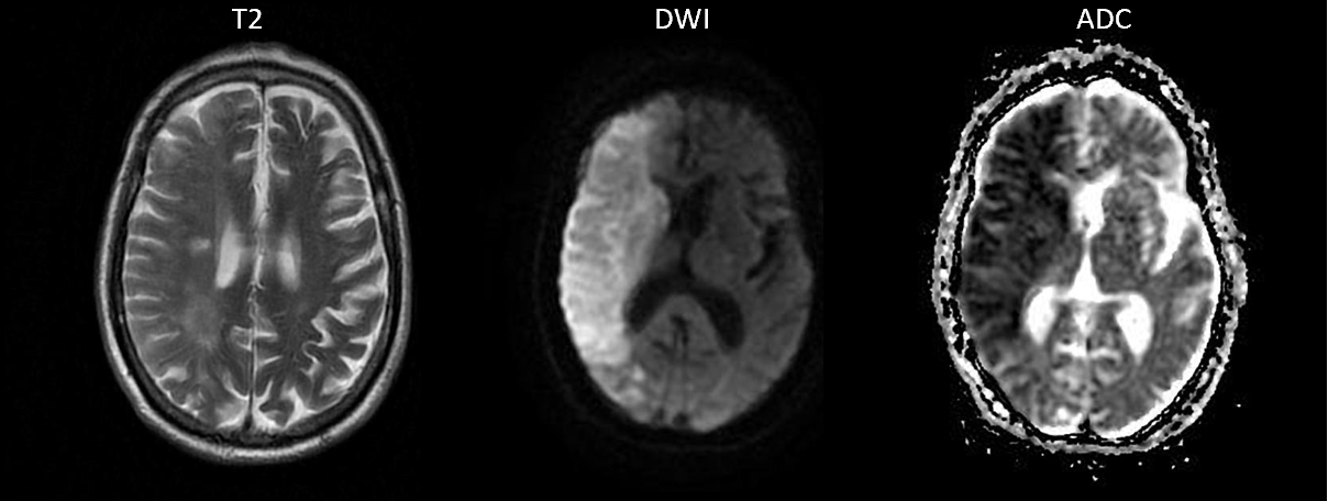
Weighted magnetic resonance imaging DW-MRI and the apparent diffusion coefficient ADC in the differentiation between benign and malignant solid head and neck masses by comparing. A b factor of zero b 0 smm 2 indicates no diffusion weighting and the image is analogous to a T2-weighted image. High b-values with or without non-Gaussian models have been used for early evaluation of cancer treatment.
In general in healthy. Using a Gaussian model with b-values up to 4000 smm 2 Mardor et al. Diffusion-weighted MRI and optimal b-value for characterization of liver lesions.
Conventional MRI and multi- b -value DW images were collected before treatment at mid-stage evaluation and when conducting therapeutic efficiency assessment after chemotherapy. The b value is used in MRI in the context of Diffusion Weighted Imaging DWI. These can either be calculated directly from the.
For example Feng et al. In general approximately 1000 smm 2 is the maximal b value for DWI 5 11. Studies have reported that the use of b values higher than 1000 smm 2 and 2000 smm 2 improves tumor.
DWI is done to determine the rate of molecular diffusion in different areas of the body.

Diffusion Tensor Imaging Dti Fiber Tracking Imagilys

Principles Of Diffusion Tensor Imaging And Its Applications To Basic Neuroscience Research Neuron
2

Diffusion Weighted Imaging Radiology Reference Article Radiopaedia Org

Principles Of Diffusion Tensor Imaging And Its Applications To Basic Neuroscience Research Neuron

Apparent Diffusion Coefficient Radiology Reference Article Radiopaedia Org

Pulse Sequences For Diffusion Weighted Mri Sciencedirect

Brain Mri Obtained From A Sagittal Plane B Axial Plane And C Download Scientific Diagram
Tensor Valued Diffusion Encoding For Diffusional Variance Decomposition Divide Technical Feasibility In Clinical Mri Systems Plos One
Signal Intensity Of Dwi And Adc In Diffusion Restriction Increased Download Scientific Diagram
Apparent Diffusion Coefficient Radiology Reference Article Radiopaedia Org
2
Diffusion Weighted Imaging Radiology Reference Article Radiopaedia Org
Diffusion Weighted Imaging Radiology Reference Article Radiopaedia Org
The Basics Of Mri Interpretation Radiology Geeky Medics
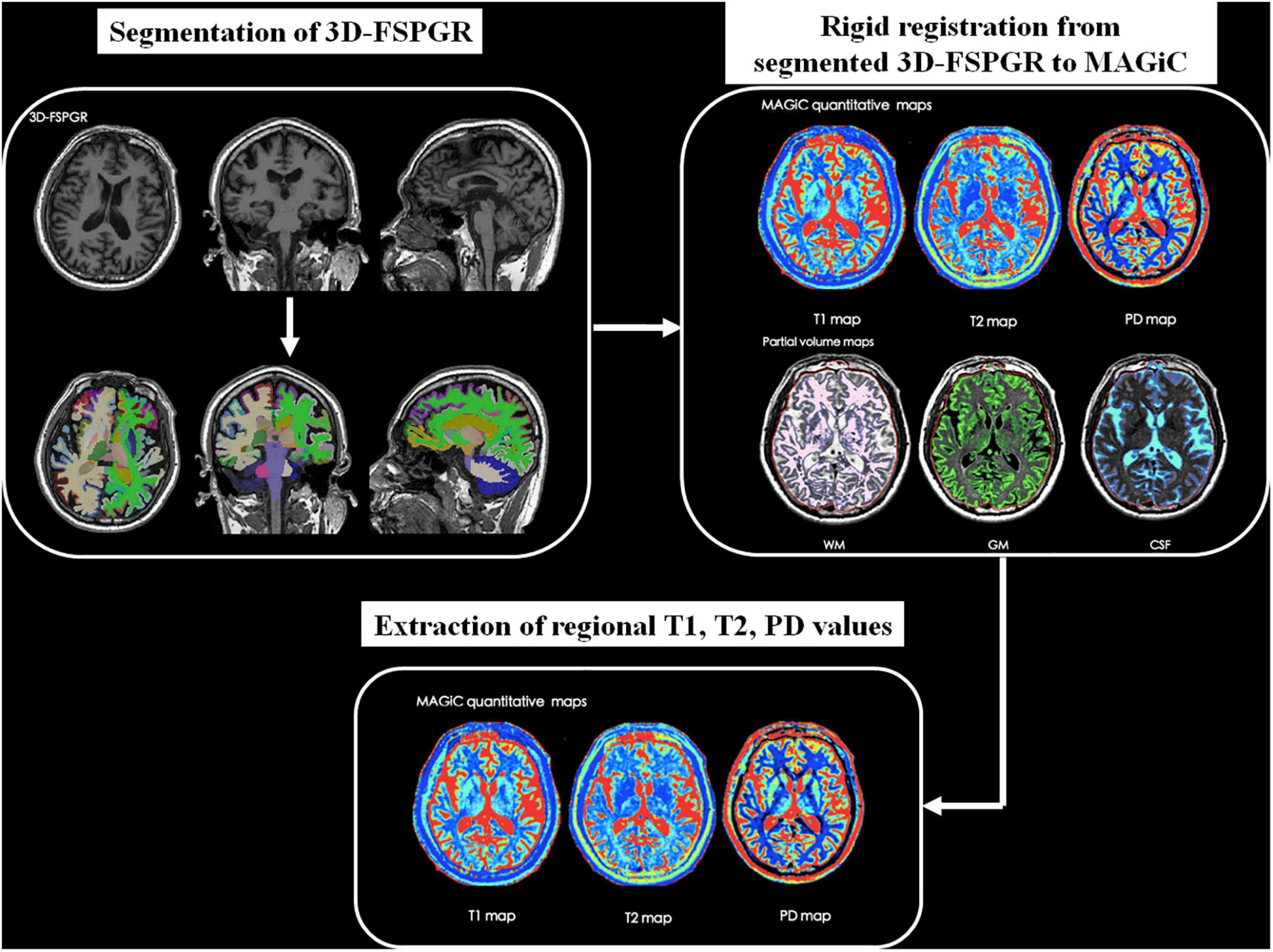
Frontiers Quantitative Analysis Of Synthetic Magnetic Resonance Imaging In Alzheimer S Disease

Hyperpolarised 13c Mri Identifies The Emergence Of A Glycolytic Cell Population Within Intermediate Risk Human Prostate Cancer Nature Communications
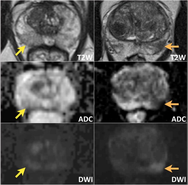
The Radiology Assistant Prostate Cancer Pi Rads V2

Apparent Diffusion Coefficient Radiology Reference Article Radiopaedia Org


Comments
Post a Comment